|
|
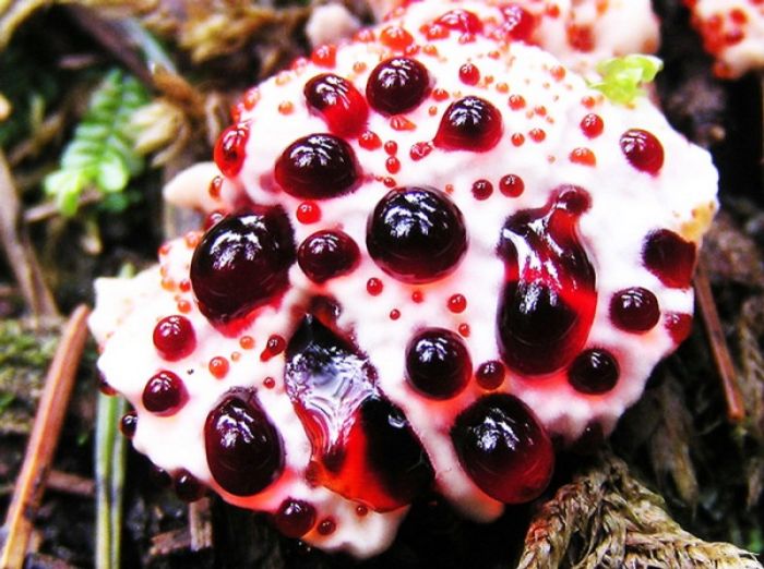
Hydnellum Peckii Fungus
|
• Microscopic features
In deposit, the spores appear brown. Viewing them with a light microscope reveals finer details of their structure: they are roughly spherical but end abruptly in a small point, their surfaces are covered with small, wart-like nodules, and their size is between 5.0–5.3 by 4.0–4.7 µm. The spores are in amyloid, meaning they do not absorb iodine when stained with Melzer's reagent.
Hydnellum peckii's cells (the hyphae) also present various characters useful for its characterization. The hyphae that form the cap are hyaline (translucent), smooth, thin-walled, and 3–4 µm thick. They collapse when dry, but may be readily revived with a weak (2%) solution of potassium hydroxide. Those in the cap form an intricate tangle with a tendency to run longitudinally. They are divided into cellular compartments (septa) and have clamp connections—short branches connecting one cell to the previous cell to allow passage of the products of nuclear division. The basidia, the spore-bearing cells in the hymenium, are club-shaped, four-spored, and measure 35–40 by 4.7–6 µm.
|
|